|
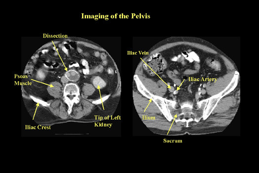
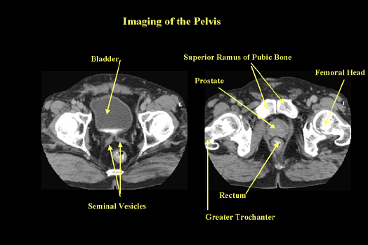
CT is excellent for imaging the soft tissues and bone in the pelvis (Figure 23 & 24). Patient preparation is important and oral contrast is usually given 1 to 2 hours before scanning to help enhance and define organ boundaries. Intravenous contrast is also routinely given and in some cases rectal contrast is administered.
|

Imaging of the Pelvis (Figure 23) |
|
The image on the left shows a dissection of the aorta. Note the oral contrast in the bowel. The image on the right shows some of the vasculature of the pelvis. The iliac artery on the left has calcified plaque on the lateral wall.
|

Imaging of the Pelvis (Figure 24) |
|
CT is an excellent modality for visualizing bony abnormalities. Further discussion on this topic will occur in the section "The Musculoskeletal System".
|
The small intestine is the largest organ in the abdomen. The 1st part is the duodenum (10 inches), the 2nd part is the jejunum (8 feet) and the3rd part is the ileum (12 feet). The function of the small intestine is absorption of foodstuffs. The large intestine is 4 feet long and is a continuation of the small intestine. Its main function is absorption of water and sodium. Arterial supply is via the superior mesenteric artery to the right side and via the inferior mesenteric artery to the left.
|